![]() Figure 7 of
Govindarajan, Mol Vis 2005;
11:876-886.
Figure 7 of
Govindarajan, Mol Vis 2005;
11:876-886.
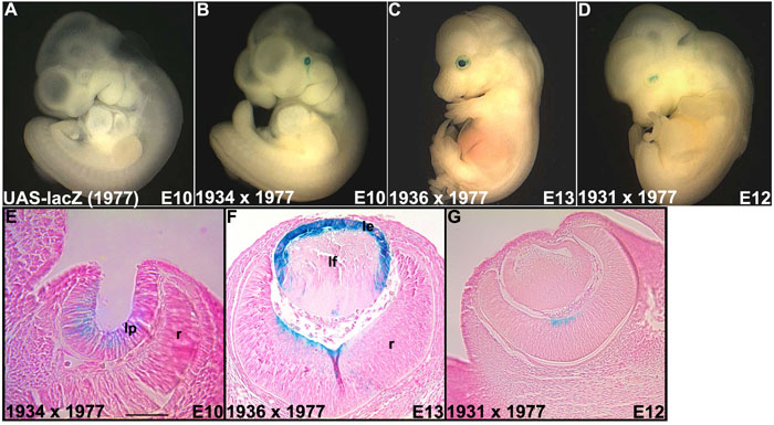
Figure 7. LacZ expression in monogenic and bigenic embryos
Embryos (E10-E13) were stained by incubation overnight in X-gal (A-D). Representative embryos were embedded, sectioned, and counterstained with nuclear fast red (E-G). No blue staining tissues were detected in monogenic UAS-lacZ embryos of the family OVE1977 (A). When the UAS-lacZ (OVE1977) mice were crossed with Pax6-GAL4/VP16 (OVE1934 [E], OVE1936 [F], and OVE1931 [G]) mice, lacZ expression was detected in the lens pit (E), lens epithelial cells (F), and retinal neuroblasts and ganglion cells (F,G). The lens epithelial cells (le), lens fiber cells (lf), lens pit (lp), and retina (r) are identified. The scale bar (in E) represents 100 μm for E-G.