![]() Figure 6 of
Govindarajan, Mol Vis 2005;
11:876-886.
Figure 6 of
Govindarajan, Mol Vis 2005;
11:876-886.
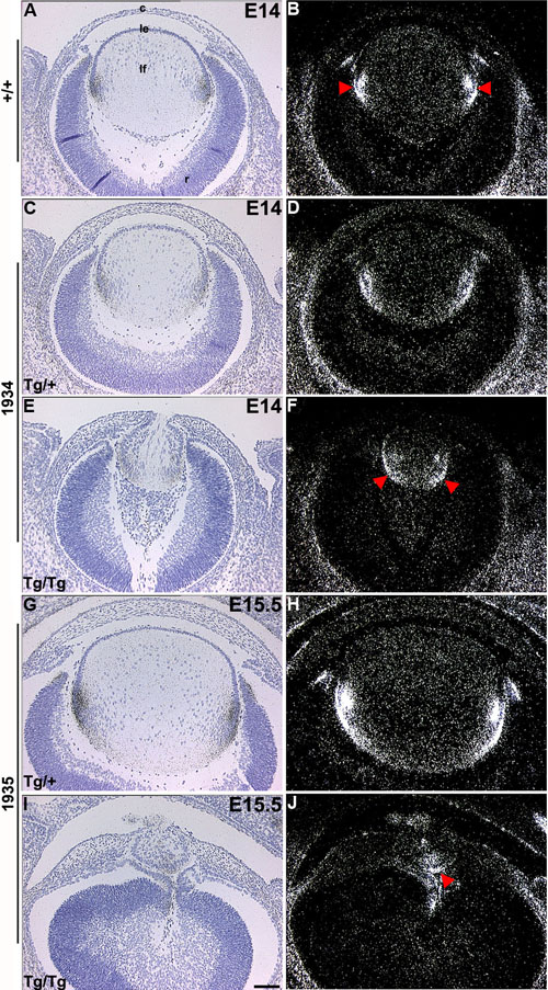
Figure 6. Expression pattern of p57Kip2
In situ hybridizations were done on ocular sections of nontransgenic (marked +/+; A,B), OVE1934 (C-F), and OVE1935 (G-J) embryos using 35S-labeled p5735 riboprobe. In wild-type lenses, p57Kip2 expression is upregulated in the equatorial region where fiber cell differentiation is initiated (A,B, red arrowheads). In lenses of homozygous (Tg/Tg) transgenic mice from OVE1934 and OVE1935 embryos, the zone of p57Kip2 induction has migrated toward the posterior of the lens (E,F,I,J, red arrowheads). Expression of p57 was nearly normal in heterozygous (Tg/+) embryos from these same families (C,D,G,H). The cornea (c), lens epithelial cells (le), lens fiber cells (lf), and retina (r) are identified in A. The scale bar (I) represents 100 μm.