![]() Figure 3 of
Govindarajan, Mol Vis 2005;
11:876-886.
Figure 3 of
Govindarajan, Mol Vis 2005;
11:876-886.
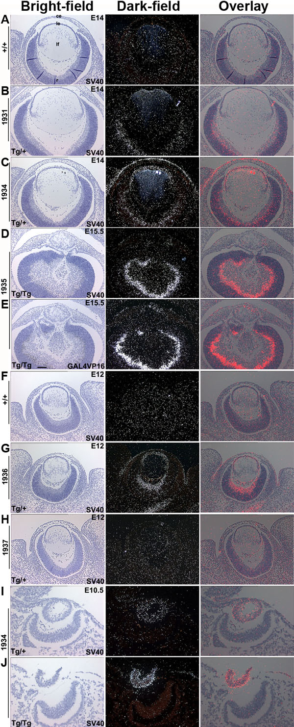
Figure 3. Transgene expression
In situ hybridizations were done on ocular sections of Pax6-GAL4/VP16 transgenic mice using 35S-labeled SV40 or GAL4/VP16 riboprobes. Bright-field, dark-field, and overlay (silver grains were pseudocolored red and overlaid on the respective bright-field image) images of ocular sections from nontransgenic (marked +/+; A,F), OVE1931 (B), OVE1934 (C,I,J), OVE1935 (D,E), OVE1936 (G), and OVE1937 (H) mice are shown. Transgene expression was detected in the lens and corneal epithelial cells in transgenic lines OVE1934, OVE1935, and OVE1936. Transgene expression was seen in a subset of retinal neuroblasts in the families OVE1931, OVE1934, OVE1935, and OVE1936. Transgene expression was also seen in some perioptic mesenchymal cells of the OVE1936 line. No transgene expression was seen in the OVE1937 line. At E10.5, homozygous (Tg/Tg) OVE1934 embryos (I) show higher levels of transgene expression relative to heterozygous (Tg/+) embryos (J). The corneal epithelium (ce), lens epithelial cells (le), lens fiber cells (lf), and retina (r) are identified in A. The scale bar (E) represents 100 μm in A-H and 50 μm in I-J.