![]() Figure 2 of
Govindarajan, Mol Vis 2005;
11:876-886.
Figure 2 of
Govindarajan, Mol Vis 2005;
11:876-886.
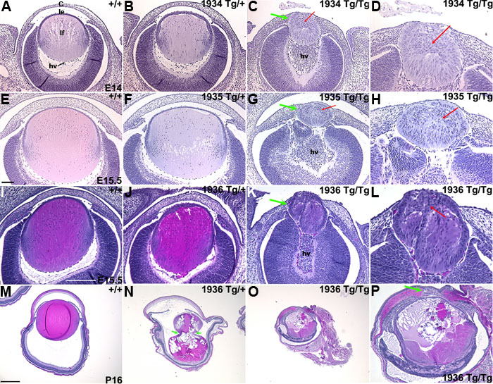
Figure 2. Altered development of the lens and cornea
Heads or eyes of nontransgenic (A,E,I,M) mice (marked +/+) and GAL4/VP16 transgenic mice were sectioned and stained with hematoxylin and eosin. Eyes of heterozygous embryos (marked Tg/+) from families OVE1934 and OVE1935 were nearly normal (B,F). Heterozygous OVE1936 embryos (E15.5) showed alterations of the lens epithelium (J). By P16, the OVE1936 mice showed cataracts with ruptured lens capsules (N). Homozygous embryos (marked Tg/Tg) from OVE1934, OVE1935, and OVE1936 families showed dramatic changes in lenticular and corneal development (C,D,G,H,K,L). The lenses in these embryos were embedded within the corneas (C,G,K, green arrows). The lens epithelial cells were stratified and disorganized (D,H,L, red arrows). The corneal stromal cells were not properly aligned in these families and development of the hyaloid vasculature (marked hv; C,G,K) was disrupted. The folded lens (M) is an artifact of the histological processing. The cornea (c), lens epithelial cells (le), and lens fiber cells (lf), are identified in A. Images in A-H and I-P were captured using two different microscopes. The scale bars in E, I, and M apply to A-H, I-L, and M-P, respectively. Scale bars represent 50 μm in D,H,L; 100 μm in A-C,E-G,I-K,P; and 250 μm in M-O. Note that C,G,K,O are shown at higher magnifications in D,H,L,P, respectively.