![]() Figure 9 of
Warejcka, Mol Vis 2005;
11:859-868.
Figure 9 of
Warejcka, Mol Vis 2005;
11:859-868.
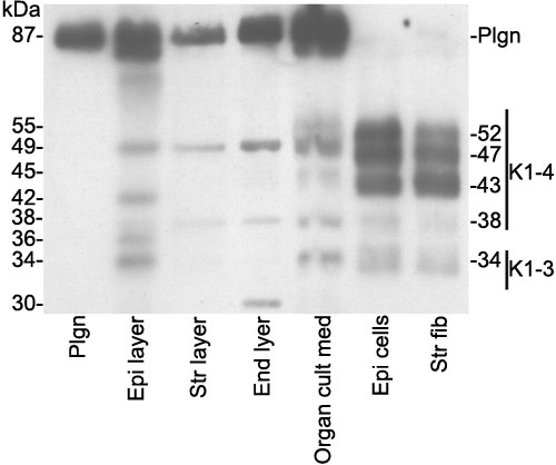
Figure 9. Comparison of angiostatins produced by cultured cells, corneal extracts, and organ culture
Corneal extracts from the epithelial (Epi), stromal (Str), and endothelial (End) layers and organ culture, epithelial (Epi) cell and stromal fibroblast (Str fib) conditioned medium were separated by SDS-PAGE and transferred to a nitrocellulose membrane. The blot was probed using two anti K1-3 angiostatin antibodies. The bands at 87 kDa are plasminogen (Plgn).