![]() Figure 8 of
Warejcka, Mol Vis 2005;
11:859-868.
Figure 8 of
Warejcka, Mol Vis 2005;
11:859-868.
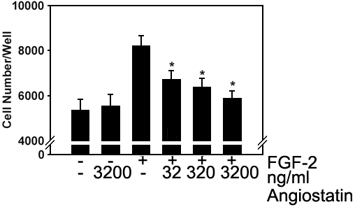
Figure 8. Stromal cell produced angiostatins inhibit proliferation of HUVECs
Plasminogen was incubated with corneal stromal fibroblasts for 96 h and angiostatin molecules were isolated on a Lysine-Sepharose column. Proliferating human umbilical vascular endothelial cells were cultured in high-glucose-DMEM with 1% L-glutamine and 5% FBS with or without FGF-2 at 60 pM and in the presence or absence of increasing amounts of cornea produced angiostatin. The cells were cultured for 72 h and the number of cells was quantified using the CYQUANT proliferation assay. Experiments we performed in triplicate. The asterisk indicates a statistically significant difference compared to FGF-2 without added angiostatin (p<0.01).