![]() Figure 5 of
Warejcka, Mol Vis 2005;
11:859-868.
Figure 5 of
Warejcka, Mol Vis 2005;
11:859-868.
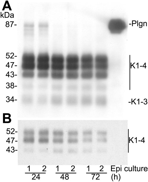
Figure 5. Human corneal epithelial cells convert plasminogen to angiostatin
A: Primary cultures of epithelial cells digested from human corneas using dispase were incubated with serum free media containing plasminogen (Plgn) for three days. At 24, 48, and 72 h, supernatant samples of two separate cultures were collected, separated by SDS-PAGE, transblotted to nitrocellulose membranes, and probed with two monoclonal antibodies to the K1-3 portion of the plasminogen molecule. B: Shorter exposure of the K1-4 angiostatins of A. After 24 h, there was very little full-length plasminogen and other products similar in size to K1-3 and K1-4 were accumulating in the supernatant. After 48 and 72 h, no full length plasminogen was detectable by western blot, but angiostatin-like molecules were easily detected. No degradation of plasminogen was observed in the control incubated for 72 h in the absence of cells.