![]() Figure 3 of
Devi, Mol Vis 2005;
11:846-852.
Figure 3 of
Devi, Mol Vis 2005;
11:846-852.
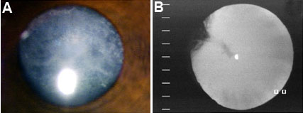
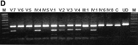
Figure 3. Analysis of Family 2
A: Photograph of the anterior eye with lens image of individual V:2, showing anterior capsular cataract with posterior cortical opacities. B: Retroillumination of the lens of individual V:1. showing peripheral cortical opacity. C: Pedigree of family 2. The shaded circle (female) and shaded boxes (males) represent the affected members and the unshaded boxes and circles denote the unaffected members in the family. The slash mark through the box/circle indicates deceased. An arrow indicates the proband. D: Restriction fragment length analysis showing a gain of an NlaIII site in the mutant that results in a 341 bp, 222 bp, 119 bp, 40 bp, and 2 bp fragments. The wild type displays 341 bp, 40 bp, and a 2 bp fragment. The 40 bp and 2 bp bands are not visible on the agarose gel.