![]() Figure 2 of
Dahlman, Mol Vis 2005;
11:88-96.
Figure 2 of
Dahlman, Mol Vis 2005;
11:88-96.
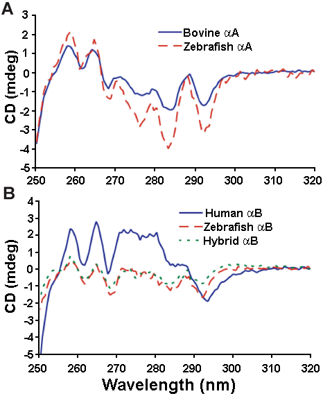
Figure 2. Near UV CD spectroscopy of mammalian and zebrafish α-crystallin
Deflections from zero millidegrees (mdeg) indicate the position of aromatic amino acids in the tertiary structure of the proteins. Shown are zebrafish and bovine αA-crystallin (A) and zebrafish, hybrid, and human αB-crystallin (B). The negative shift in the zebrafish and hybrid αB-crystallin spectra between 270 and 290 nm is likely due to the addition of two tryptophans and six tyrosines compared to the human ortholog. All scans were performed at 22 °C.