![]() Figure 1 of
Dahlman, Mol Vis 2005;
11:88-96.
Figure 1 of
Dahlman, Mol Vis 2005;
11:88-96.
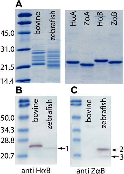
Figure 1. SDS-PAGE and western blot analysis of bovine and zebrafish total lens soluble proteins
A: Coomasie blue stained 12% SDS-PAGE gel of 3 μg total lens soluble protein from bovine and zebrafish lens and 1 μg of each of the following recombinant proteins; human αA-crystallin (HαA), zebrafish αA-crystallin (ZαA), human αB-crystallin (HαB), and zebrafish αB-crystallin (ZαB). B: Western blot of bovine and zebrafish total lens soluble proteins probed with an antibody to human αB-crystallin. C: Western blot of bovine and zebrafish total lens soluble protein probed with an antibody to zebrafish αB-crystallin. Arrow 1 indicates a weak zebrafish αB-crystallin band recognized by the antibody to human αB-crystallin. Arrow 2 indicates two closely grouped zebrafish αA- and αB-crystallin bands recognized by the antibody to zebrafish αB-crystallin. Arrow 3 indicates a third band of unknown identity recognized by the same antibody. Molecular sizes of standards are indicated in kilodaltons to the left of each gel.