![]() Figure 4 of
Ohno-Matsui, Mol Vis 2005;
11:1-10.
Figure 4 of
Ohno-Matsui, Mol Vis 2005;
11:1-10.
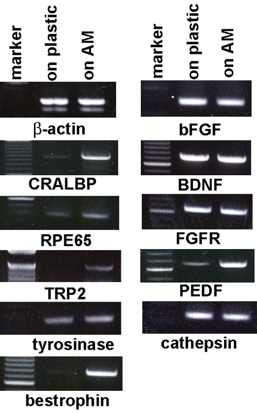
Figure 4. RT-PCR analysis of discriminatory molecules and growth and trophic factors in RPE cells
Total RNA was extracted from confluent RPE cells cultured on either plastic or amniotic membrane (AM). RT-PCR was performed on 2 μg RNA. For each gel shown, the left lane (labeled "marker") is a commercially supplied size marker, the middle lane (labeled "on plastic") is product from a reaction using RNA from RPE cells cultured on plastic, and the right lane (labeled "on AM") is a product from a reaction using RNA from RPE cells cultured on AM. cDNA product (2 μl) from RT-PCRs that used β-actin primers was used for all PCR reactions and β-actin was used as a positive control throughout.