![]() Figure 3 of
Ohno-Matsui, Mol Vis 2005;
11:1-10.
Figure 3 of
Ohno-Matsui, Mol Vis 2005;
11:1-10.
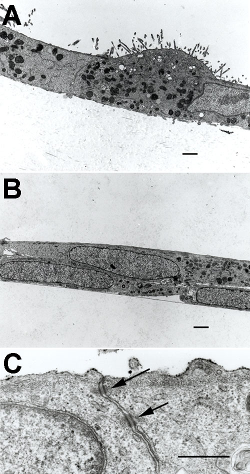
Figure 3. Transmission electron micrographs of cultivated RPE cells on denuded amniotic membrane for two weeks
A: Electron micrographs show a tight monolayer of cuboidal to spheroidal RPE cells growing over amoniotic membrane (AM). A heterogenous expression of apical microvilli was observed on the apical side. B: RPE cells cultured on plastic show elongated cell shape in multilayers. C: Electron micrographs reveal junctional specializations on the apical side between adjacent cells cultured on AM. Scale bars represent 1 μm.