![]() Figure 2 of
Ohno-Matsui, Mol Vis 2005;
11:1-10.
Figure 2 of
Ohno-Matsui, Mol Vis 2005;
11:1-10.
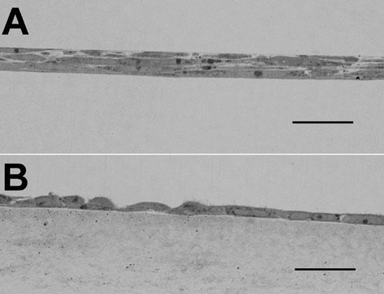
Figure 2. Different morphology of RPE cells cultured on plastic and on amniotic membrane
Toluidine blue stained sections of RPE cells on plastic (A) and RPE cells after 14 days cultivation on amniotic membrane (B). Scale bars represent 50 μm.