![]() Figure 1 of
Ohno-Matsui, Mol Vis 2005;
11:1-10.
Figure 1 of
Ohno-Matsui, Mol Vis 2005;
11:1-10.
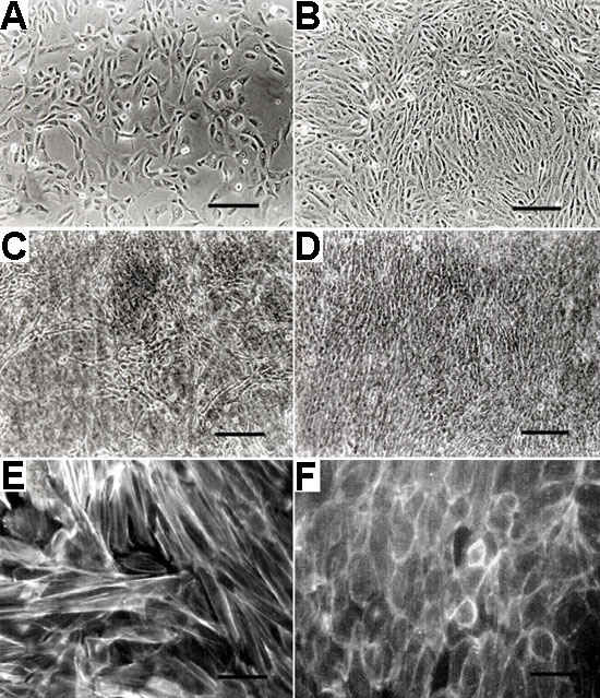
Figure 1. Phase contrast micrographs and F-actin immunostaining
Human RPE cells cultured on plastic one day after seeding (A) and at confluence (B). Human RPE cells cultured on amniotic membrane (AM) one day after seeding (C) and at confluence (D). F-actin immunostaining in confluent RPE cells on plastic (E) and on AM (F). Scale bars in A-D represent 100 μm; scale bars in E and F represent 15 μm.