![]() Figure 5 of
Larsson, Mol Vis 2004;
10:821-831.
Figure 5 of
Larsson, Mol Vis 2004;
10:821-831.
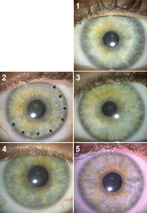
Figure 5. Contraction Furrows
This scale measured the extension and distinction of contraction furrows in the main stroma leaf of the iris. Photo 1 in the scale had no contraction furrows. In photo 2, a contraction furrow extends halfway from the periphery of the iris from 1 to 8 o clock. In photo 3, the contraction furrows are more distinct and extend around the whole iris. In photo 4, the contraction furrows are more distinct. In photo 5, a minimum of two full circles of contraction furrows present. A corresponding scale showing what the contraction furrows look like in brown colored iris were also used to assist the rater judgments.