![]() Figure 4 of
Larsson, Mol Vis 2004;
10:821-831.
Figure 4 of
Larsson, Mol Vis 2004;
10:821-831.
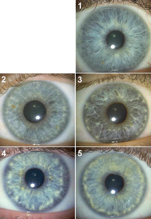
Figure 4. Wolfflin Nodules
Photo 1 in the scale has no Wolfflin nodules at the periphery of the iris. In photo 2, a ring of clumped nodules extends from 3 to 10 o'clock. In photo 3, the white nodules are more visible and distinct from each other. In photo 4, the nodules lager, are lighter and are closer together. In photo 5, the Wolfflin nodules have formed a uniform white ring that is much thicker and distinct than in the previous scale steps. A corresponding scale, showing what the Wolfflin nodules look like when partly covered by pigment was used to assist the raters judgments.