![]() Figure 2 of
Fujii, Mol Vis 2004;
10:814-820.
Figure 2 of
Fujii, Mol Vis 2004;
10:814-820.
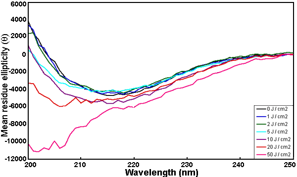
Figure 2. Far UV CD spectrum of normal and UV-C irradiated α-crystallin
Change in secondary structure of α-crystallin by UV-C irradiation was measured using far UV CD. The protein concentration was 0.1 mg/ml in 50 mM Tris-HCl buffer at pH 7.8 and the cell path length was 0.2 mm. The secondary structure of α-crystallin was altered with increasing doses of UV-C irradiation.