![]() Figure 6 of
Frederikse, Mol Vis 2004;
10:794-804.
Figure 6 of
Frederikse, Mol Vis 2004;
10:794-804.
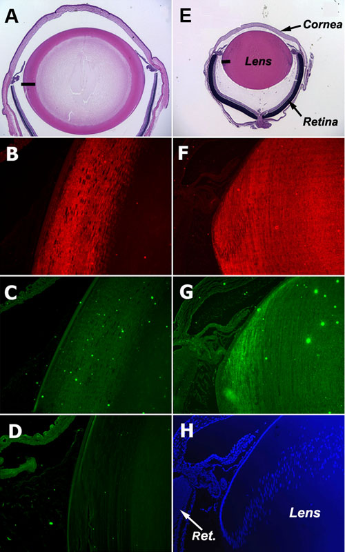
Figure 6. Analysis of site 1 phosphorylated synapsin protein distribution in post-natal and adult lenses
Analysis of site-1 phosphorylated synapsin and site-1 dephosphorylated synapsin protein in adult (A-D) and post-natal day 5 (E-H) rat lenses. A,E: Hematoxylin and Eosin histological stain. B,F: anti-site-1 dephosphosynapsin specific antibodies. C,G: anti-site-1 phosphosynapsin specific antibodies. D: primary antibody omitted. H: DAPI nuclear stain. Black horizontal bars in A and E indicate the general area shown in B-D and F-H, respectively.