![]() Figure 3 of
Yin, Mol Vis 2004;
10:787-793.
Figure 3 of
Yin, Mol Vis 2004;
10:787-793.
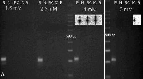
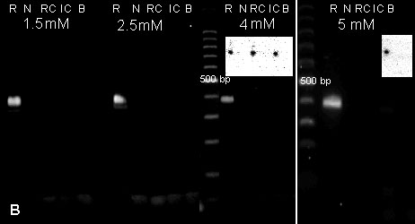
Figure 3. M1 receptor mRNA expressed in rat but not chick tissue: PCR and Southern analyses
PCR and Southern analyses of chick and rat mRNA suggested M1 receptor expression was confined to rat tissue alone. PCR amplification with primers M1a (A) and M1b (B) at an annealing temperature of 58 °C. Four different MgCl2concentrations, 1.5 mM, 2.5 mM, 4 mM, and 5 mM, were used. The tissue samples used in each MgCl2 titration were rat brain cDNA (R), negative control (N), chick retina-choroid complex (RC), iris-ciliary body complex (IC) and brain (B). DNA fragments of the expected size (see Table 1 for details) were observed in rat positive controls (207 and 385 bp for primer pairs M1a and M1b, respectively). There were no fragments of the expected size in any of the chick samples. Southern blot and probing with 32P radiolabeled M1 sequence (insets) confirmed the PCR findings.