![]() Figure 2 of
Yin, Mol Vis 2004;
10:787-793.
Figure 2 of
Yin, Mol Vis 2004;
10:787-793.
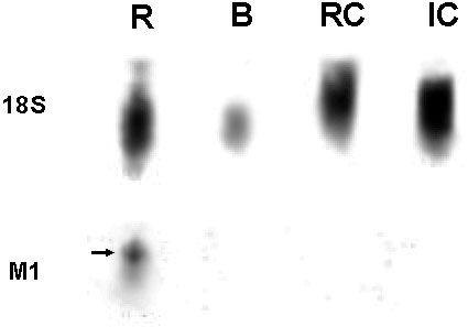
Figure 2. M1 receptor mRNA expressed in rat but not chick tissue: Northern analysis
Northern analysis of chick and rat mRNA suggested M1 receptor expression was confined to rat tissue alone. The northern membrane labeled with the 18S probe (top picture) showed a strong positive signal for all samples (rat brain: R, chick brain: B, retina-choroid complex: RC, and iris-ciliary body complex: IC; 30 μg of total RNA). The membrane labeled with the M1 probe (bottom picture) only showed hybridization to the rat sample (R). The arrow indicates the specific binding region. No chick sample (B, RC, or IC) showed any specific hybridization.