![]() Figure 2 of
Li, Mol Vis 2004;
10:750-757.
Figure 2 of
Li, Mol Vis 2004;
10:750-757.
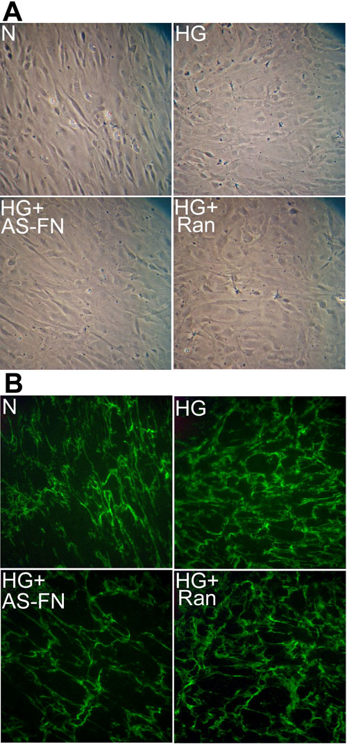
Figure 2. FN immunoreactivity in HTM cells
A: Phase contrast micrographs shows HTM cells grown in normal medium (N), high glucose medium (HG), high glucose medium transfected with antisense FN oligo (HG+AS-FN), or random oligo (HG+Ran). The magnification was 100x. B: Representative photomicrographs shows increased FN immunostaining in cells grown in high glucose medium compared to cells grown in normal medium. Specific reduction in FN immunostaining was observed in cells transfected with AS-FN oligos. C: Graphical illustration of relative FN immunoreactivity in cells grown in normal medium, high glucose medium or cells grown in high glucose medium and transfected with either AS-FN oligos or random oligos. Data is expressed as mean±standard deviation; the asterisk indicates a p value of <0.05 (n=6).