![]() Figure 7 of
White, Mol Vis 2004;
10:738-749.
Figure 7 of
White, Mol Vis 2004;
10:738-749.
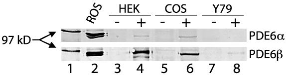
Figure 7. Adenovirus expression of rod PDE6α and PDE6β subunits
HEK293, COS7, and Y79 cells were transduced with adenovirus engineered to express PDE6α or PDE6β subunits, followed by western analysis of cell extracts using a PDE6 specific antibody. Equal amounts of cytosol extracted proteins (about 80 mg) were loaded into each lane. Two bands migrating close to the 97 kDa marker (lane 1) corresponding to the α and β subunits are observed in bovine ROS (lane 2). In the upper panel PDE6α subunit protein is detectable in trace amounts in HEK293 (lane 4) and COS7 transduced cells, but is not observed in Y79 cells (lane 8). In the lower panel PDE6β is readily detectable in HEK and COS cells and barely detectable in Y79 cells. No bands are observed at the corresponding molecular weights in non-transduced cells (lanes 3, 5, and 7).