![]() Figure 6 of
White, Mol Vis 2004;
10:738-749.
Figure 6 of
White, Mol Vis 2004;
10:738-749.
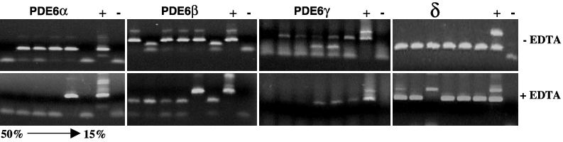
Figure 6. Polysome profile of PDE6 transcripts in Y79 cells
Polysomes were isolated from the Y79 cell line using a sucrose gradient in the presence or absence of EDTA. RNA in each polysome fraction was amplified with primers specific for PDE subunit mRNA to determine the distribution in the gradient. For each primer set the lane order from left to right is six lanes of gradient fractions from 50 to 15% sucrose, and positive (+) and negative control (-) without template. In the upper panels for all primer sets transcripts are evenly distributed across fractions indicating that the mRNA is in a complex with ribosomes or other proteins. An A260 profile of the fractionated gradient demonstrated intact polysomes (data not shown). Addition of EDTA (lower panels) dissociates mRNA from the ribosome and shifts the transcripts to the less dense sucrose fractions. PDE6α, β, γ shows a marked shift to free RNA, however δ transcripts remained in all fractions analyzed indicating that δ mRNA may be bound to other proteins that could inhibit translation.