![]() Figure 3 of
White, Mol Vis 2004;
10:738-749.
Figure 3 of
White, Mol Vis 2004;
10:738-749.
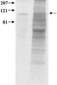
Figure 3. Metabolic labeling and immunoprecipitation of Y79 cell protein
Y79 proteins were metabolically labeled (right lane) with 35S-Met followed by immunoprecipitation of total protein cell extract with a PDE6 specific polyclonal antibody (left lane). A single polypeptide of 99 kDa (arrow pointing left) corresponding with the size of the α and β subunits was absorbed by the antibody. Molecular mass markers, in kDa, are shown to the left of the gel image.