![]() Figure 1 of
Liu, Mol Vis 2004;
10:712-719.
Figure 1 of
Liu, Mol Vis 2004;
10:712-719.
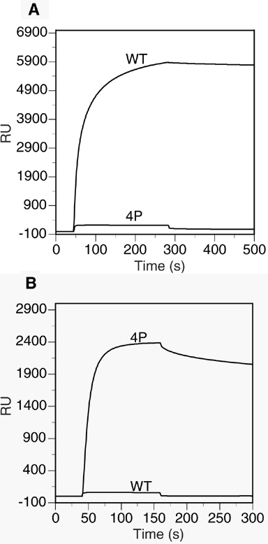
Figure 1. The binding of anti-rhodopsin antibodies to WT and 4P peptides
The peptides, WT and 4P, were immobilized on a CM5 sensor chip at approximately 400 resonance units (RU) using procedures described in Methods. A: 333 nM 1D4 antibody, which recognizes the unphosphorylated carboxyl terminus of rhodopsin, was injected onto the sensor chip from 40-280 s, followed by continuous flow of buffer alone beginning at 280 s to measure the dissociation phase. The level of binding was measured as a change in resonance units (RU). B: 333 nM A11-82P, which recognizes the phosphorylated carboxyl terminus of rhodopsin, was measured for binding to the sensor chip as described in A, except that antibody was injected from 40-160 s, followed by continuous flow of buffer alone beginning at 160 s to measure the dissociation phase. The data are representative of two independent experiments that gave very similar results.
