![]() Figure 6 of
Nakamura, Mol Vis 2004;
10:703-711.
Figure 6 of
Nakamura, Mol Vis 2004;
10:703-711.
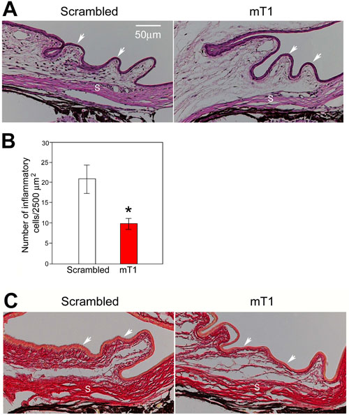
Figure 6. Suppression of inflammatory response and collagen deposition by TβRII siRNA
A: Hematoxylin and eosin stained tissue sections from mouse eyes 2 days after injection with PBS/latex beads along with 200 nM scrambled or mT1 siRNA. Arrows indicate the conjunctiva and S indicates the sclera. The bar represents 50 μm. B: Bar graph showing the number of inflammatory cells per 2,500 μm2 of subconjunctival area in eyes injected with 200 nM of mT1 compared to those treated with scrambled siRNA. Data are presented as means (n=5); error bars represent the standard error of the mean. The asterisk indicates values significantly different from those of scrambled RNA treated controls (p<0.015). C: Tissue sections from eyes 14 days after injection were stained with picrosirius red to demonstrate deposition of collagen that appears as pink fibrillar structures. Note the thick subconjunctival collagen layers in the scrambled siRNA treated mouse eye. Arrows indicate the conjunctival epithelium and S indicates the sclera.