![]() Figure 1 of
Nakamura, Mol Vis 2004;
10:703-711.
Figure 1 of
Nakamura, Mol Vis 2004;
10:703-711.
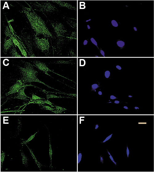
Figure 1. siRNA inhibits TβRII expression
Immunofluorescence analysis of human corneal fibroblasts, untreated (A,B), treated for 48 h with scrambled siRNA (C,D), or with 100 nM hT1 (E,F) was performed. TβRII expression is shown in A,C, and E. Staining of nuclei with DAPI (blue) is shown in B, D, and F. Note the reduction in TβRII staining of cells treated with hT1 siRNA (E) compared to control cells (A,C). The morphology after the siRNA treatment was somewhat altered. The bar represents 10 μm.