![]() Figure 3 of
Lin, Mol Vis 2004;
10:688-695.
Figure 3 of
Lin, Mol Vis 2004;
10:688-695.
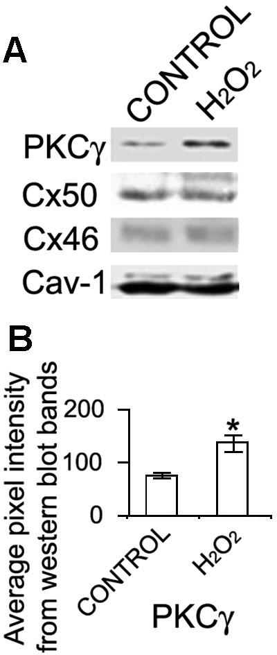
Figure 3. Coimmunoprecipitation of PKCγ with Cav-1, Cx46, or Cx50 in lipid rafts
Whole lenses were treated with 100 μM H2O2 for 15 min. Whole cell extracts were isolated and subjected to sucrose gradient centrifugation. Lipid raft enriched fractions 3 to 6 were pooled and immunoprecipitated with anti-Cav-1. The immunoprecipitated Cav-1 protein complexes were analyzed by western blot using anti-Cav-1, PKCγ, Cx46, or Cx50 (A). Cav-1 was present as a loading control. The intensity of the PKCγ bands was digitalized and graphed (B). The experiments were carried out in triplicate and representative blots are shown. The asterisk indicates statistical significance.