![]() Figure 2 of
Lin, Mol Vis 2004;
10:688-695.
Figure 2 of
Lin, Mol Vis 2004;
10:688-695.
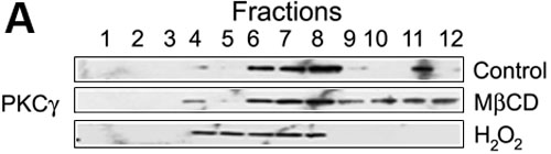
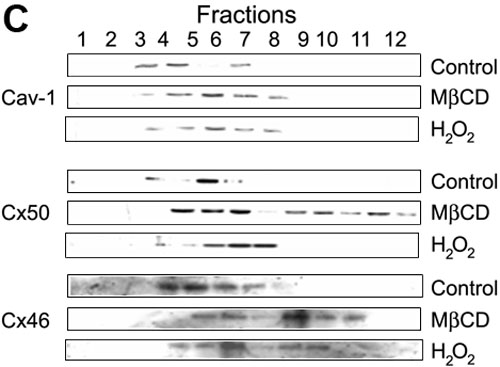
Figure 2. Distribution of PKCγ, Cav-1, Cx50, and Cx46 in lipid rafts
Rat lens whole cell extracts with or without H2O2 treatment were isolated and fractionated on 5-35% sucrose continuous density gradients as described in Methods. Fractions (1 ml each) were collected from the top of each gradient. Proteins were precipitated by 10% TCA, separated by SDS-PAGE, and immuno-visualized by western blotting. Fractions 3 to 6 were considered as lipid raft fractions containing caveolin-1 (Cav-1) [22]. A: Fractionation of PKCγ localization patterns. The PKCγ patterns were graphed by western blot band intensity and shown in B. Results shown are the means of three experiments. The asterisk indicates statistical significance. C: Fractionation of Cav-1, Cx50, and Cx46 localization patterns. MβCD is an abbreviation for methyl-β-cyclodextrin and IB abbreviates immunoblot.