![]() Figure 1 of
Lin, Mol Vis 2004;
10:688-695.
Figure 1 of
Lin, Mol Vis 2004;
10:688-695.
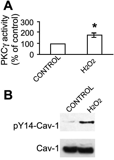
Figure 1. PKCγ activation by H2O2 in the rat lens
A: Rat lenses (six weeks of age) were treated with 100 μM H2O2 for 15 min. Whole cell extracts were isolated and endogenous PKCγ was immunoprecipitated with specific anti-PKCγ antisera and its activity was measured by use of the PepTag PKC assay kit. Enzyme activity was normalized by calibration of the relative level of phosphorylated substrates to the relative amount of PKCγ in the immunoprecipitation as determined by western blotting, and was expressed as percent of untreated specific PKCγ activity. The experiments were carried out in triplicate. The error bars represent the standard error of the mean for the three samples. The asterisk indicates statistical significance. B: Phosphorylation of caveolin-1 (Cav-1) on Tyr14 is stimulated by H2O2. Lens whole cell extracts were fractionated on 5-35% sucrose continuous density gradients as described in the Methods section. Lipid rafts enriched fractions 3 to 6 were pooled and immunoprecipitated. Total proteins were resolved by SDS-PAGE and by western blot with anti-phospho-Tyr14 on Cav-1 (pY14-Cav-1) and anti-Cav-1. H2O2 dramatically stimulated Cav-1 on Tyr14. This is as a positive control for initiation of a H2O2 stress response in the whole lens.