![]() Figure 3 of
McKay, Mol Vis 2004;
10:682-687.
Figure 3 of
McKay, Mol Vis 2004;
10:682-687.
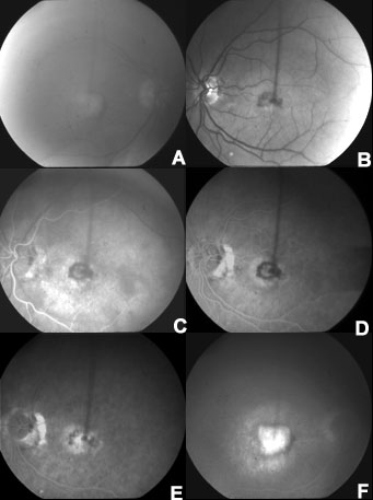
Figure 3. Clinical phenotype of AMD patient with hemicentin Gln5345Arg mutation
Red free image of right eye with a disciform scar and significant lens opacities (A) and left eye with a subfoveal choroidal neovascular membrane (B). Fluorescein angiogram of left eye at 25 s with early filling of a subfoveal choroidal neovascular membrane (C), 57 s (D), and 3 min (E). Late stage fluorescein of the right eye with a disciform scar (F).