![]() Figure 2 of
McKay, Mol Vis 2004;
10:682-687.
Figure 2 of
McKay, Mol Vis 2004;
10:682-687.
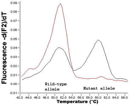
Figure 2. Differentiation of mutant and wild type alleles following PCR and melting curve analysis
The peaks indicate the temperatures at which rapid decreases in fluorescence occur. These represent the melting temperatures of the sensor probes for the wild type (51 °C) and mutant alleles (60 °C). As the probes melt off their target they are no longer adjacent to the anchor probe, Fluorescence Resonance Energy Transfer (FRET) can therefore no longer take place and the fluorescence emitted drops. The red line is a homozygous wild type sample with a large peak at the melting temperature of the wild type probe. The blue line is a heterozygote, with a decreased wild type peak and an additional peak indicating presence of the mutant allele.