![]() Figure 8 of
Elias, Mol Vis 2004;
10:672-681.
Figure 8 of
Elias, Mol Vis 2004;
10:672-681.
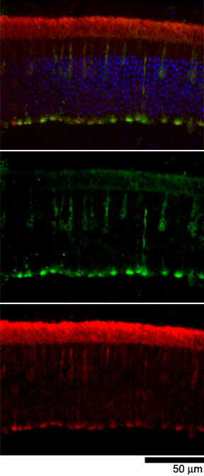
Figure 8. Co-labeling of cones with anti mouse cone arrestin and anti-Arrestin
Co-labeling of cones with anti mouse cone arrestin and anti-Arrestin. Double labeling of a section of a retina from a light-adapted mouse (top panel) demonstrates the co-labeling of cones with rabbit anti mouse cone arrestin (green) and mouse MH785 anti-arrestin (red) and provides independent support of the conclusion that MH785 labels rod and cone arrestin. The same section viewed with a filter to detect mouse cone arrestin alone (middle row) and arrestin alone (bottom row) are also shown. The scale bar represents 50 μm.