![]() Figure 3 of
Elias, Mol Vis 2004;
10:672-681.
Figure 3 of
Elias, Mol Vis 2004;
10:672-681.
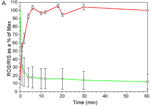
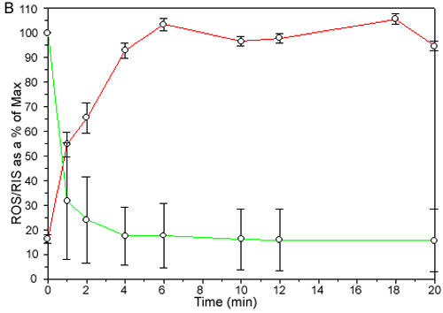
Figure 3. Temporal kinetics of dark to light translocation of arrestin and rod α-T
A: Temporal kinetics of dark to light translocation of arrestin and rod α-T. Grayscale immunofluorescent images were quantified by scanning densitometry using Metamorph Image Analysis software and the ROS/RIS ratios determined. The Arrestin graph in red indicates ROS/RIS as a percentage of ROS/RIS at 60 min versus the specified time. The Transducin graph in green indicates ROS/RIS as a percentage of ROS/RIS at 0 min versus the specified time. Each point in the time course experiments is from a single animal and represents the average values (the error bars represent the standard deviation) from three different fields within a section. These data are representative of those obtained from three separate experiments. B: The same data are shown on an expanded time scale. The ROS/RIS ratio for α-T reaches its minimum value by 1.5 min whereas arrestin reaches its maximum value in about 4.5 min.