![]() Figure 4 of
Ledee, Mol Vis 2004;
10:663-667.
Figure 4 of
Ledee, Mol Vis 2004;
10:663-667.
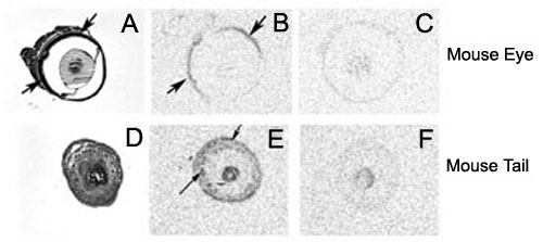
Figure 4. In situ hybridization of a riboprobe directed against the full length CCL27 chemokine mRNA
A: Hematoxilin and eosin staining of a section of the mouse eye used for hybridization studies. B: Film autoradiogram showing specific hybridization of the full length CCL27 antisense riboprobe in the retina (arrows) in the same section. Similar results were obtained with antisense riboprobes directed against intron 1, exon 3, and the PESKY exon 1'. C: Film autoradiogram of a consecutive section hybridized with sense (control) riboprobe. D: Hematoxylin-eosin staining of a section of the mouse tail used for hybridization studies. E: Film autoradiogram showing CCL27 mRNA expression in the skin (arrows) in the same section. F: Film autoradiogram of a consecutive section hybridized with sense (control) riboprobe.