![]() Figure 2 of
Pasta, Mol Vis 2004;
10:655-662.
Figure 2 of
Pasta, Mol Vis 2004;
10:655-662.
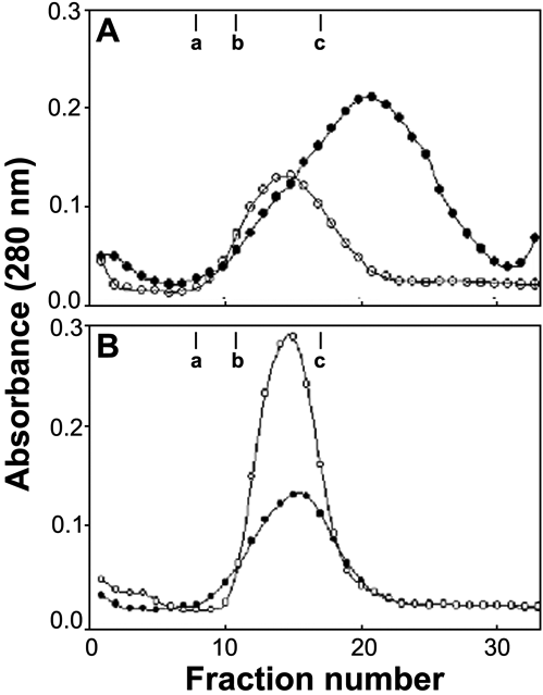
Figure 2. Glycerol density gradient centrifugation of wild type α-crystallins and the gxg mutants
The proteins were sedimented through a linear 10-40% gradient of glycerol. A: αA-crystallin (open circle) and αAgxg-crystallin (filled circle). B: αB-crystallin (open circle) and αBgxg-crystallin (filled circle). The positions of proteins used for standard molecular masses are also indicated. The marker band at "a" indicates aldolase (158 kDa), the marker band at "b" indicates catalase (232 kDa), and the marker band at "c" indicates thyroglobulin (669 kDa).