![]() Figure 3 of
Hackam, Mol Vis 2004;
10:637-649.
Figure 3 of
Hackam, Mol Vis 2004;
10:637-649.
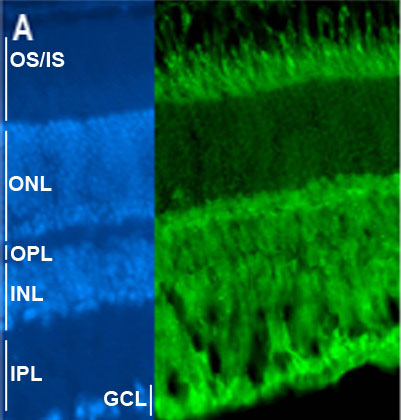
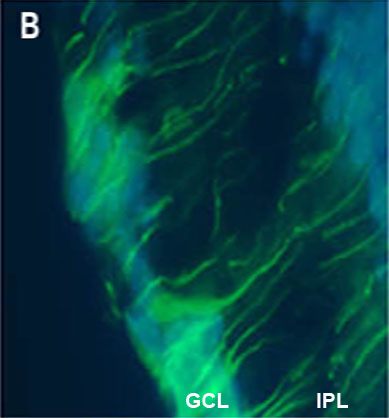
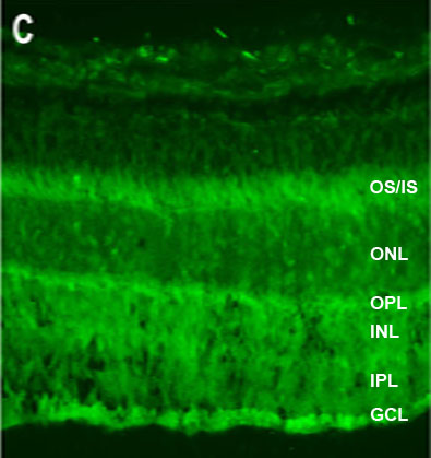
Figure 3. The cellular localization of two retina enriched RNA binding proteins, EWS and PCPB1
The nuclei have been counterstained using DAPI (blue). EWS appears cytoplasmic in each cell layer (A, green), and is prominent in cytoplasmic extensions that may be Müller glia processes (B, green). PCPB1 is expressed in all cell layers (C, green), with higher staining intensity in the ganglion cell layer (GCL), inner segments (IS), and the outer edge of the inner nuclear layer (INL). The outer nuclear layer (ONL), outer segments (OS), outer plexiform layer (OPL), and inner plexiform layer (IPL) are also labeled.