![]() Figure 2 of
Hackam, Mol Vis 2004;
10:637-649.
Figure 2 of
Hackam, Mol Vis 2004;
10:637-649.
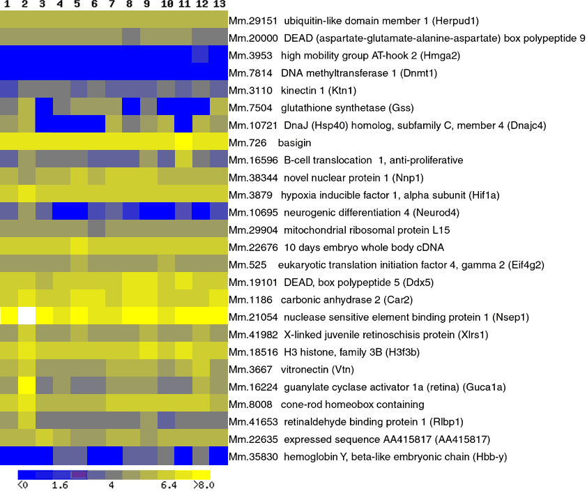
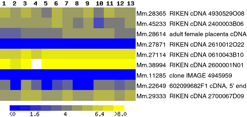
Figure 2. Regional distribution of retina-enriched genes in the CNS
The regional distribution in the CNS of a subset of annotated genes (A) and ESTs (B), determined by comparison to publicly available Affymetrix oligonucleotide microarray data from 13 different neuronal regions. 1 hippocampus, 2 eye, 3 trigeminal, 4 hypothalamus, 5 spinal cord lower, 6 spinal cord upper, 7 amygdala, 8 striatum, 9 olfactory bulb, 10 DRG, 11 cerebellum, 12 cortex, 13 frontal cortex. The expression distribution is visualized by assigning colors to the log of the average signal intensity values. Original negative values were assigned the lowest color value (blue), the remainder were assigned to ten different colors based on their log values. White represents the highest log values. The color key indicates the range of expression levels represented by each color.