![]() Figure 4 of
Stoilov, Mol Vis 2004;
10:629-636.
Figure 4 of
Stoilov, Mol Vis 2004;
10:629-636.
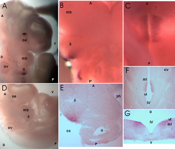
Figure 4. Details of Cyp1b1 expression at 10.5 dpc
A: Lateral view focused on the optic eminence (oe), otic vesicle (ov) and the developing pharyngeal region including the mandibular component of the first branchial arch (mb) and the second branchial arch (II). B: Frontal view of the developing pharyngeal region. C: Dorsal view of hindbrain focused on the floor of the fourth ventricle. D: Control embryo. E: Frontal section through the second branchial arch (II). The arch artery (a) is also labeled. F,G: Frontal and sagittal sections, respectively, through the Cyp1b1 expression domain in the hindbrain. The fourth ventricle (IV) and ventricular layer (vl) are also labeled. For orientation, anterior (A), posterior (P), dorsal (D), and ventral (V) are labeled.