![]() Figure 3 of
Stoilov, Mol Vis 2004;
10:629-636.
Figure 3 of
Stoilov, Mol Vis 2004;
10:629-636.
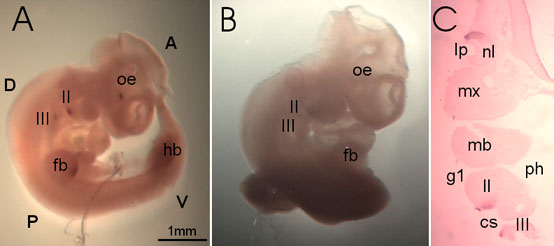
Figure 3. Cyp1b1 expression at 10.5 dpc
A: Lateral view. Cyp1b1 expression domains are now consolidated to the caudal tip of the second (II) and third (III) arch. Strong, demarcated expression domains are visible in the optic eminence (oe) and the forelimb bud (fb). A less defined signal is detected in the developing hindlimb bud (hb). B: Control embryo hybridized with sense probe. C: Frontal section through the eye and the developing branchial arches. Cyp1b1 expression is limited to the dorsal half of the inner layer of the optic cup, the future neural layer (nl) of the retina. In the branchial arches, expression is seen in both the ectoderm and the neighboring mesenchyme. The branchial arches 2-3 (II-III), lens pit (lp), lumen of the pharynx (lp), cervical sinus (cs), mandibular component of first branchial arch (mb), maxillary component of first branchial arch (mx), otic vesicle (ov), and branchial groove 1 (g1) are also labeled. For orientation, anterior (A), posterior (P), dorsal (D), and ventral (V) are labeled. The bar represents a length of 1 mm.