![]() Figure 2 of
Stoilov, Mol Vis 2004;
10:629-636.
Figure 2 of
Stoilov, Mol Vis 2004;
10:629-636.
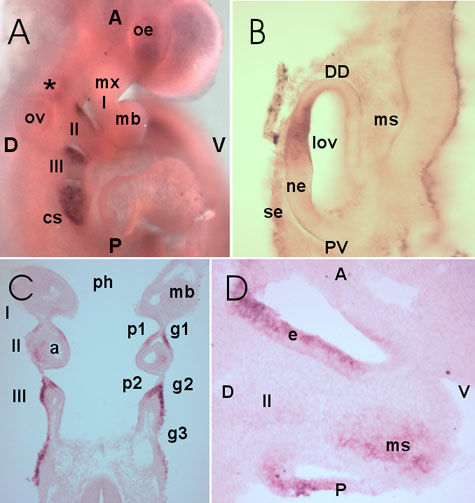
Figure 2. Details of Cyp1b1 expression at 9.5 dpc
A: Lateral view of the whole mount. B: Frontal section through the developing optic vesicle. With respect to the dorso-distal/proximo-ventral axis of the developing eye (DD/PV), Cyp1b1 expression was restricted to dorsal half of the neuroectoderm (ne) in the future inner layer of the optic cup. The mesenchyme (ms) and surface ectoderm (se) are also labeled. C: Frontal section through the developing branchial arches. The branchial arches 1-3 (I-III), branchial pouches (p1,p2), branchial grooves (g1-g3), arch arteries (a), mandibular component of first branchial arch (mb), and lumen of the pharynx (ph) are also labeled. D: Lateral section through the second branchial arch (II). Cyp1b1 expression is detected in ectoderm (e) and mesenchyme (ms) of the developing arch. The future cervical sinus (cs), lumen of the optic vesicle (lov), maxillary component of first branchial arch (mx), optic eminence (oe), otic vesicle (ov), and hindbrain expression domain (*) are also labeled. For orientation, anterior (A), posterior (P), dorsal (D), and ventral (V) are labeled.