![]() Figure 1 of
Stoilov, Mol Vis 2004;
10:629-636.
Figure 1 of
Stoilov, Mol Vis 2004;
10:629-636.
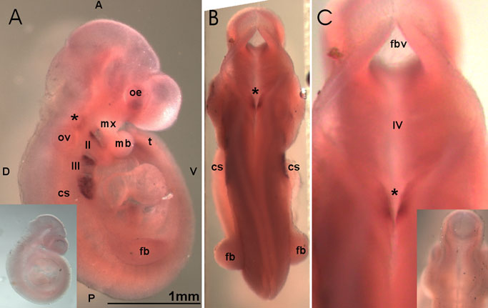
Figure 1. Cyp1b1 expression at 9.5 dpc
A: Lateral view. A strong, demarcated Cyp1b1 expression domains are seen in the second (II) and third (III) branchial (pharyngeal) arches including the area of the future cervical sinus (cs). A much weaker and less defined signal is seen in the developing forelimb bud (fb), optic eminence (oe), and the caudal end of the tail (t) where the future hindlimb bud will develop. The relative transparency of the tissue makes it possible to appreciate the location of hindbrain expression domain (*) in relation to the otic vesicle (ov). The mandibular component of first branchial arch (mb) and maxillary component of first branchial arch (mx) are also labeled. B: Dorsal view. C: Dorsal view focused on the expression domain in the hindbrain region (*). The fourth ventricle (IV) and forebrain vesicle (fvb) are also labeled. The inserts in A and C represent control embryos hybridized with sense probe. For orientation, anterior (A), posterior (P), dorsal (D), and ventral (V) are labeled. The bar represents a length of 1 mm.