![]() Figure 5 of
Fernandes, Mol Vis 2004;
10:618-628.
Figure 5 of
Fernandes, Mol Vis 2004;
10:618-628.
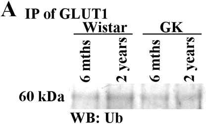
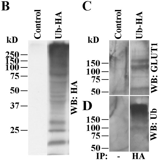
Figure 5. Evidence for ubiquitinylation of GLUT1 in vivo
A: GLUT1 was immunoprecipitated from retinal lysates obtained from Wistar (control) and GK (diabetic) rats. Proteins were resolved by SDS-PAGE, transferred to PVDF membranes, and probed with antibodies directed against ubiquitin conjugates (FK2). The additional band with molecular weight around 60 kDa is compatible with a monoubiquitinylated form of GLUT1. B: HEK cells were transfected with multiubiquitin tagged to HA (Ub-HA). The cells were lysed and their proteins were resolved by SDS-PAGE, transferred to PVDF membranes, and probed with antibodies directed against HA. This showed efficient transfection and formation of endogenous Ub-HA conjugates had occurred. C: Immunoprecipitates of Ub-HA conjugates were subsequently probed with antibodies directed against GLUT1. D: Samples were immunoprecipitated with anti-HA and probed with antibodies directed against ubiquitin conjugates.