![]() Figure 6 of
Hahn, Mol Vis 2004;
10:598-607.
Figure 6 of
Hahn, Mol Vis 2004;
10:598-607.
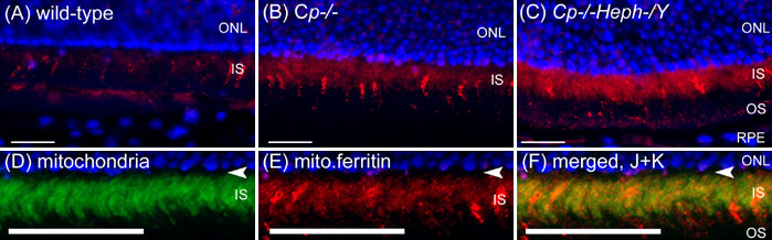
Figure 6. Mitochondrial ferritin is present in normal inner segments and is increased in Cp-/- and Cp-/-Heph-/Y retinas
Fluorescence photomicrographs of age matched wild type, Cp-/-, and Cp-/-Heph-/Y retinas immunolabeled for mitochondrial ferritin (A-C) and imaged under identical exposure parameters. Cp-/- retinas (B) have increased mitochondrial ferritin (red) in the inner segments (IS) compared to wild type (A), and Cp-/-Heph-/Y retinas (C) have further increased mitochondrial ferritin. The label in the Cp-/- inner segments (E) excludes the inner segment myoid (arrowhead, D-F) and colocalizes with a mitochondria-specific antibody (green) to the inner segment ellipsoid (D,F), suggesting mitochondrial localization of MtF. Nuclei are labeled with DAPI (blue). Scale bars represent 50 μm.