![]() Figure 5 of
Hahn, Mol Vis 2004;
10:598-607.
Figure 5 of
Hahn, Mol Vis 2004;
10:598-607.
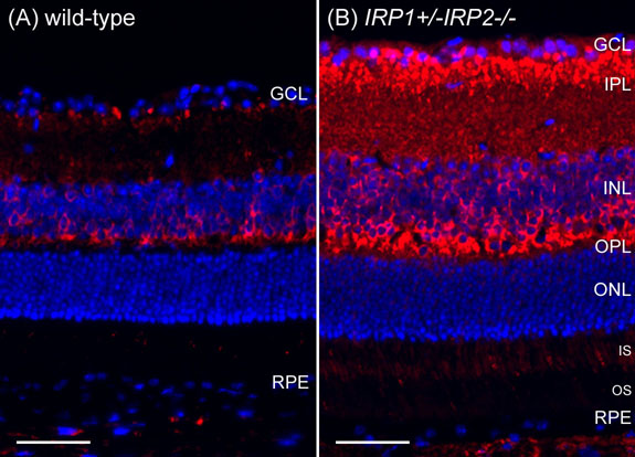
Figure 5. IRP1+/-IRP2-/- retinas have increased L-ferritin
Fluorescence photomicrographs of age matched wild type and IRP1+/-IRP2-/- retinas immunolabeled for L-ferritin (red), counterstained for nuclei with DAPI (blue), and imaged under identical exposure parameters. L-ferritin is increased in the IRP1+/-IRP2-/- inner retina, including bipolar cell synapses in the IPL, as well as in the OPL and inner segments. Scale bars represent 50 μm.