![]() Figure 4 of
Hahn, Mol Vis 2004;
10:598-607.
Figure 4 of
Hahn, Mol Vis 2004;
10:598-607.
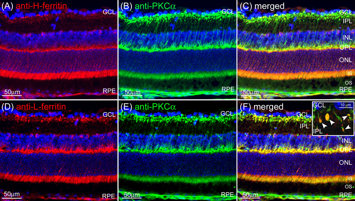
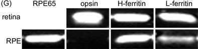
Figure 4. H- and L-ferritin are present in normal retina in rod bipolar cells
Normal retina labeled for H-ferritin (red; A) and L-ferritin (red; D). Nuclei are counterstained with DAPI (blue). Both ferritin subunits have punctate label in the inner IPL, the region adjacent to the ganglion cell layer (GCL). This punctate label is characteristic of the synaptic terminals of rod bipolar cells, labeled with anti-PKCα (green), which colocalizes (gold) with both H-ferritin (B-C) and L-ferritin (E-F). This colocalization is best seen in high power images of these synaptic terminals which are positive for both PKCα and L-ferritin (arrowheads, inset F). H- and L-ferritin mRNAs are also expressed in retina and RPE, as indicated by bands from ethidium bromide-stained agarose gels corresponding to RT-PCR amplification products of the indicated mRNAs and of the expected size from dissected C57BL/6 murine RPE and retina (G).