![]() Figure 2 of
Hahn, Mol Vis 2004;
10:598-607.
Figure 2 of
Hahn, Mol Vis 2004;
10:598-607.
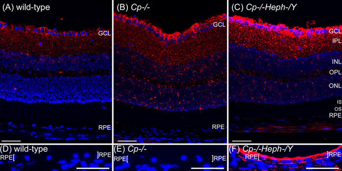
Figure 2. Cp-/-Heph-/Y retinas have increased ferroportin
Fluorescence photomicrographs of age matched wild type, Cp-/-, and Cp-/-Heph-/Y retinas immunolabeled for ferroportin (red) and imaged under identical exposure parameters. Nuclei were counterstained with DAPI (blue). Differences between wild type (A) and Cp-/- (B) were subtle, but there is a clear increase in ferroportin label in the Müller endfeet of the Cp-/-Heph-/Y retina (C). The punctate ferroportin label throughout the IPL is also increased in the Cp-/-Heph-/Y retina. In order to optimally detect differences in ferroportin in the pigmented RPE, it was necessary to pre-bleach sections (D-F). Equivalently bleached retinas immunolabeled with ferroportin and imaged with equivalent exposure parameters reveals a robust increase in the Cp-/-Heph-/Y RPE of ferroportin, which localized to both the apical and basolateral surfaces of the RPE (demarcated with brackets). Scale bars represent 50 μm.