![]() Figure 1 of
Hahn, Mol Vis 2004;
10:598-607.
Figure 1 of
Hahn, Mol Vis 2004;
10:598-607.
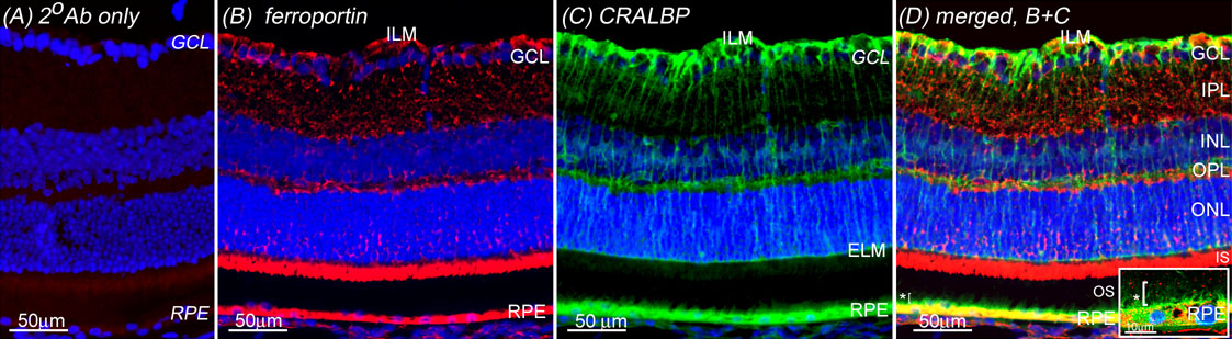
Figure 1. Ferroportin is present in normal retina at high levels in the Müller cell endfeet and RPE
Normal BALB/c retina immunolabeled with omission of primary antibody (A) has undetectable autofluorescence or background immunolabel even at longer exposure times than all other displayed panels. Nuclei are labeled with DAPI (blue). Ferroportin label (red) in normal BALB/c retina (B) localizes to Müller endfeet near the ILM, in photoreceptor inner segments (IS), and in the RPE, as demonstrated by the co-label with CRALBP (green), a marker for Müller cells and RPE (C,D). Ferroportin in the RPE excludes its apical microvilli, the green only label indicated with an asterisk ("*") in D and in the inset of D, which shows a high power image of RPE co-labeled with ferroportin and CRALBP. Ferroportin is also present in a punctate pattern throughout the inner retina.