![]() Figure 6 of
Simon, Mol Vis 2004;
10:588-597.
Figure 6 of
Simon, Mol Vis 2004;
10:588-597.
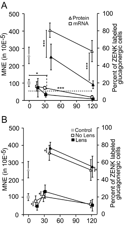
Figure 6. Comparison of ZENK protein and ZENK mRNA expression
Comparison of ZENK protein expression in retinal glucagon amacrine cells (determined by immunohistochemistry, triangles and re-plotted after Bitzer and Schaeffel [9]) to mRNA concentrations (determined by real time RT-PCR, squares) after negative lens treatment (A) and positive lens treatment (B). The scales of the two ordinates were chosen so as to avoid overlapping of the data points from both techniques. Gray symbols indicate untreated control animals, filled black symbols lens treated eyes and open symbols contralateral control eyes. Statistically significant differences between controls and the various treatment groups were determined by REST and are marked with asterisks over the horizontal lines (single asterisks p<0.05, triple asterisks p<0.001). Differences between the "No Lens" and "Lens" groups at a given time were determined by paired t test and are indicated by vertical lines with an asterisk (single asterisks p<0.017).