![]() Figure 5 of
Jablonski, Mol Vis 2004;
10:577-587.
Figure 5 of
Jablonski, Mol Vis 2004;
10:577-587.
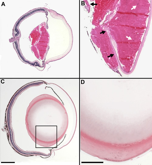
Figure 5. Comparative ocular histology of RIIIS/J and wildtype mice
Examples of the ocular histology of an RIIIS/J mouse (A and B) and a non-mutant F2 intercross progeny (C and D). Both the eye and lens of the RIIIS/J mouse are considerably smaller than that of the non-mutant mouse. In addition, histologic analysis reveals severe abnormalities in lens structure. In all RIIIS/J mice, the lens fails to form a spherical shape and lens tissue is present behind the lens capsule within vitreous chamber of the eye. Moreover, the epithelial cells circumscribe the lens (black arrows), rather than terminating at the lens equator (A and B). Nuclei fail to degrade in the lens fiber cells of the mutant mouse (white arrows). There are no apparent abnormalities in the eyes of non-mutant mice. Insets in A and C are magnified in B and D, respectively. The magnification bars are equal to 1 mm.